Short Description:
# Shoulder Joint Anatomical Model – The Precise Partner for Medical Teaching
In the field of medical teaching and clinical research, precise and intuitive anatomical models are the key cornerstones for knowledge transmission and professional improvement. This shoulder joint anatomical model is precisely a high-quality teaching aid meticulously crafted to deeply meet professional needs.
1. Precise restoration, building clear cognition
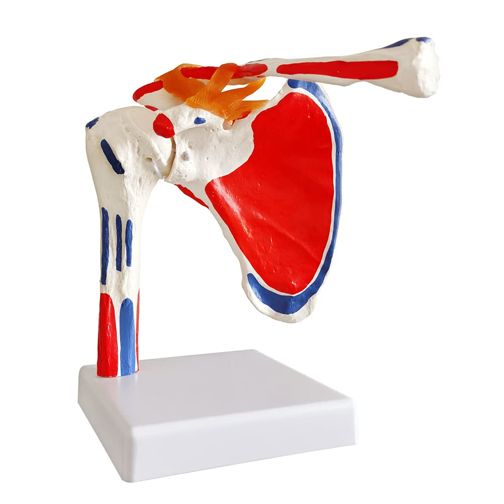
The model strictly replicates the anatomical structure of the real human shoulder joint with precision. From the fine texture of the bones, the shape of the articular surface, to the wrapping range of the joint capsule, the direction and tension of the ligaments, and even the layered distribution of the muscle tissue, all are restored to the millimeter level. The bone shapes such as the humerus and scapula are realistic. The joint capsule is clearly defined by the prominent red area, which plays a protective and restrictive role in the movement of the shoulder joint. Ligaments and muscles marked in different colors precisely present anatomical positions and connection relationships, helping learners quickly establish three-dimensional spatial cognition, break through the understanding barriers of planar graph teaching, and make the complex anatomical knowledge of the shoulder joint intuitive and accessible.
Second, diverse scenarios, adapting to professional needs
In the classrooms of medical colleges and universities, it serves as a “visual teaching plan” for teachers’ lectures. It can be flexibly disassembled and combined to dynamically demonstrate the principles of shoulder joint flexion, extension, rotation and other movements, transforming abstract anatomical knowledge into concrete operations to help students consolidate their foundation. In clinical scenarios, doctors conduct preoperative planning with the help of models, which can clearly simulate the impact of lesions on the structure of the shoulder joint and precisely formulate surgical plans. When facing patients, the model is used to visually display the connection between the patient’s condition and anatomy, enabling patients to immediately understand the logic of diagnosis and treatment, and significantly improving communication efficiency and compliance.
Third, excellent quality ensures long-term use
Made of high-quality, environmentally friendly and medical-grade materials, it combines durability and safety. It can withstand repeated disassembly and assembly as well as touching. It is not prone to deformation or fading after long-term use and does not emit any odor, providing stable and reliable tool support for teaching and clinical scenarios. Whether it is cultivating professional talents in medical education or assisting precision medicine in clinical diagnosis and treatment, this shoulder joint anatomy model, with its professional strength, has become a solid support for exploring the mysteries of the shoulder joint and enhancing professional understanding, helping every user to delve deeper into the medical field.