Short Description:
This model is suitable for use as an intuitive teaching aid when teaching physiological hygiene courses in ordinary middle schools, enabling students to understand the distribution of bronchioles in the lungs and their division into terminal bronchioles, as well as their relationship with alveoli.
# Alveolar Anatomical Model – The “Microscopic Window” for Respiratory System Teaching
Want to directly unravel the mysteries of alveoli and respiratory physiology? This “Alveolar Anatomy Model” builds an accurate bridge for medical teaching and biological science popularization, taking you through the core position of gas exchange!
1. Precise Restoration, “Visualization” of Anatomical Structures
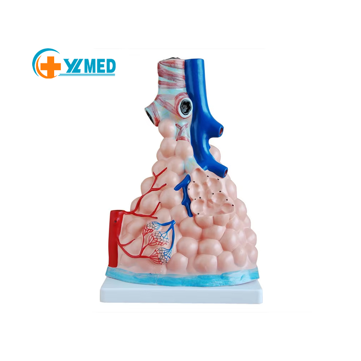
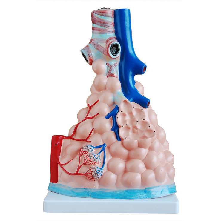
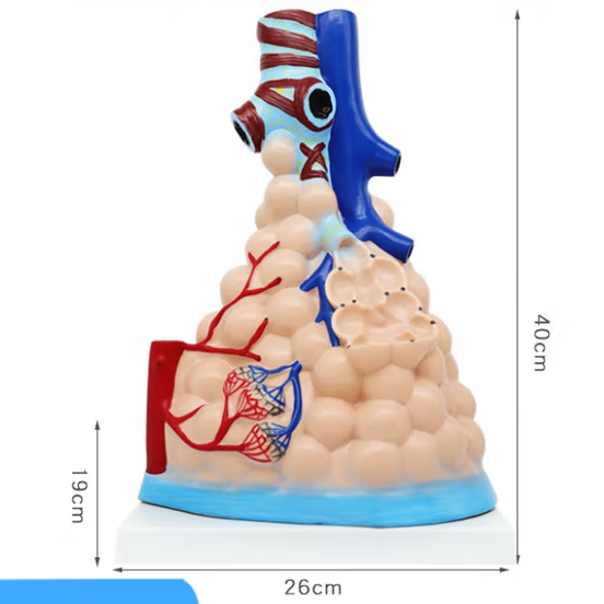
The model presents the associated structure of alveoli and bronchioles in a ** high simulation proportion ** completely:
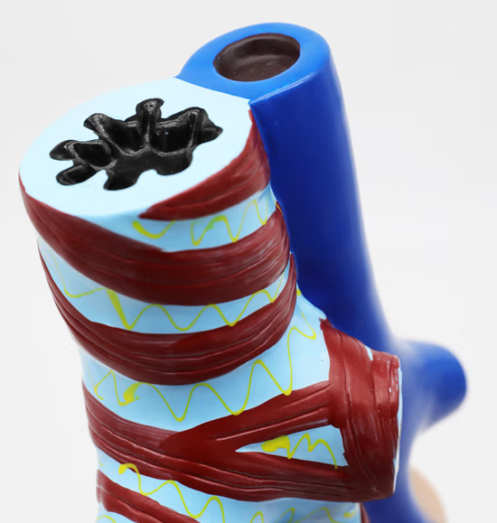
- ** Airway System ** : Clearly display the hierarchical branches of terminal bronchioles → respiratory bronchioles → alveolar ducts → alveolar sacs, restore the “tree-like network” of the airway, and help you understand the gas delivery path;
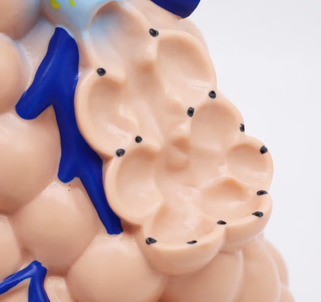
- ** Alveolar Unit ** : Magnifies and presents the morphology of alveoli, as well as the microscopic structures such as the capillary network and elastic fibers within the alveolar septum, providing an intuitive explanation of the “structural basis of gas exchange” – how oxygen passes through the alveolar walls and capillary walls into the blood, and how carbon dioxide is expelled in the opposite direction;
- ** Vascular Distribution ** : Mark the connections between the pulmonary artery, branches of the pulmonary vein and capillaries, clearly presenting the specific operation of the “pulmonary circulation” in the alveoli, and cracking the collaborative logic of the respiratory and circulatory systems.
Second, multi-scenario usage to make knowledge “within easy reach”
(1) Medical Education: The Transition from Theory to Practice
- ** Classroom Teaching ** : Teachers can combine models to explain knowledge such as “the role of alveolar surfactant” and “Changes in alveolar structure during emphysema”, replacing abstract descriptions with “physical” demonstrations to make respiratory physiology and pathology knowledge easier to understand.
- ** Student Practical Operation ** : Medical students can strengthen their memory of key points such as “qi-blood barrier” and “alveolar ventilation-blood flow ratio” by recognizing the model structure, laying a foundation for the study of “Physiology”, “Pathology”, and “Internal Medicine”.
(2) Biological Science Popularization: Making Breathing Knowledge “Vivid”
- ** Campus Science Popularization ** : In middle school biology classes, models are used to demonstrate questions such as “Why does breathing become rapid after running?” (the demand for alveolar ventilation increases) and “How does smoking harm alveoli?” (it destroys the elastic fibers of alveoli), making the abstract principle of breathing intuitive and interesting;
- ** Public Health Promotion ** : In community health lectures and hospital science popularization exhibition halls, models are used to explain the pathogenesis of “chronic obstructive pulmonary disease and pneumonia”, helping the public understand the essence of diseases and enhance their awareness of health protection.
(3) Clinical Training: Assisting in understanding respiratory diseases
- ** Nurse/Rehabilitation Therapist Training ** : By observing the model, understand “how nebulization therapy drugs reach the alveoli” and “how chest physical therapy promotes alveolar ventilation”, and optimize nursing and rehabilitation operations;
- ** Patient Education ** : Doctors can visually demonstrate the “structural changes after alveolar injury” to patients with chronic obstructive pulmonary disease and pulmonary fibrosis, assist in explaining treatment plans (such as pulmonary rehabilitation training and drug targets), and enhance patient compliance.
Three, high-quality design, durable and realistic
Made of ** environmentally friendly PVC material **, it features a stable structure, high color reproduction, and can be used for a long time without deformation. The base design ensures that the model can be placed stably, facilitating multi-angle observation and explanation. Whether it is high-frequency teaching demonstrations or long-term display displays, it can accurately convey knowledge and become a “permanent teaching aid” for respiratory physiology learning.
From the theoretical classes of medical students to public health science popularization, this alveolar anatomy model, with its intuitive “microscopic perspective”, makes breathing knowledge no longer obscure!
Teaching content:
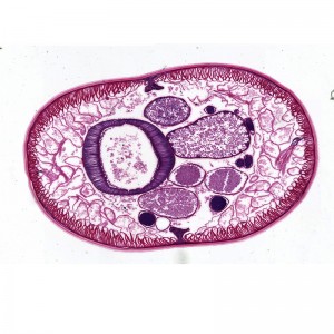
1. Cross-section of cartilageless bronchioles;
2. The relationship between terminal bronchioles and alveoli;
3. The structure of alveolar ducts and alveolar sacs;
4. The capillary network contained in the compartments between alveoli.
Made of PVC and placed on a plastic base. Dimensions: 26x15x35CM.
Packaging: 81x41x29CM, 4 pieces per box, 8KG