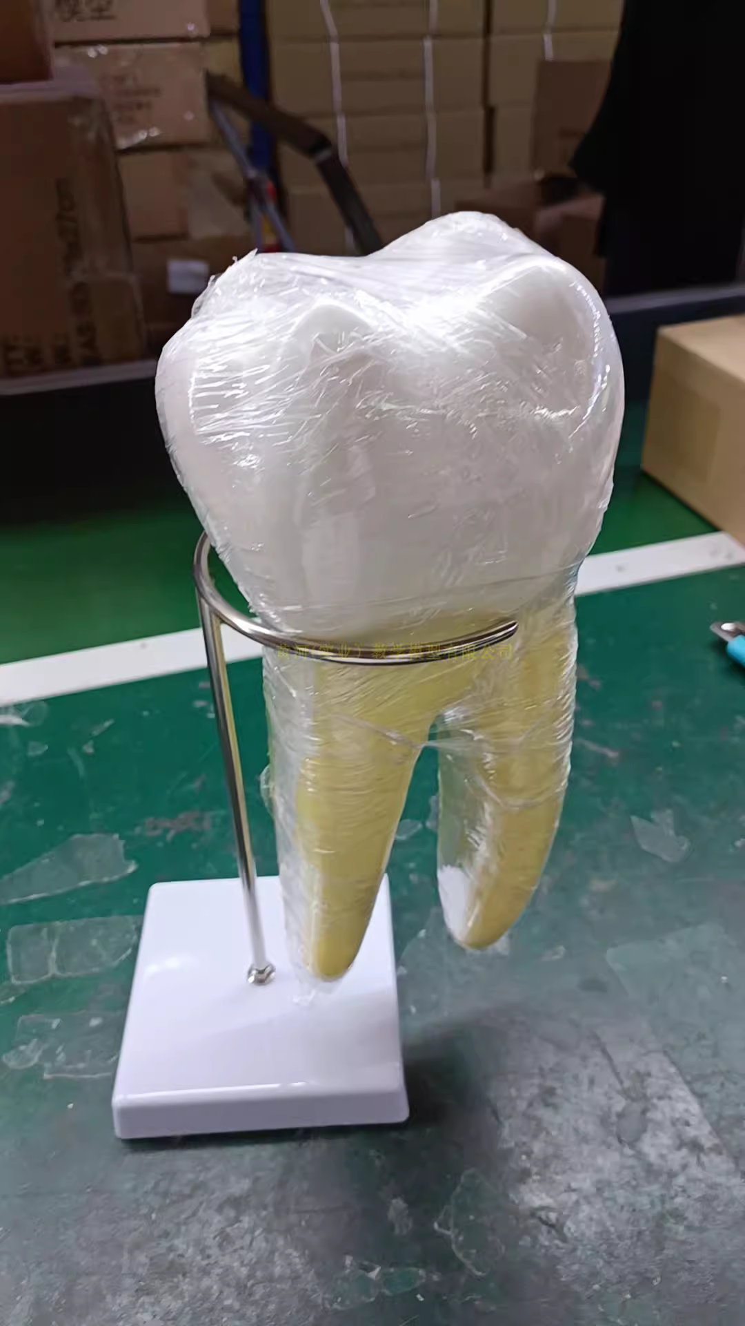
Human molars anatomy model, kindergarten teeth teaching AIDS, large teeth model with base
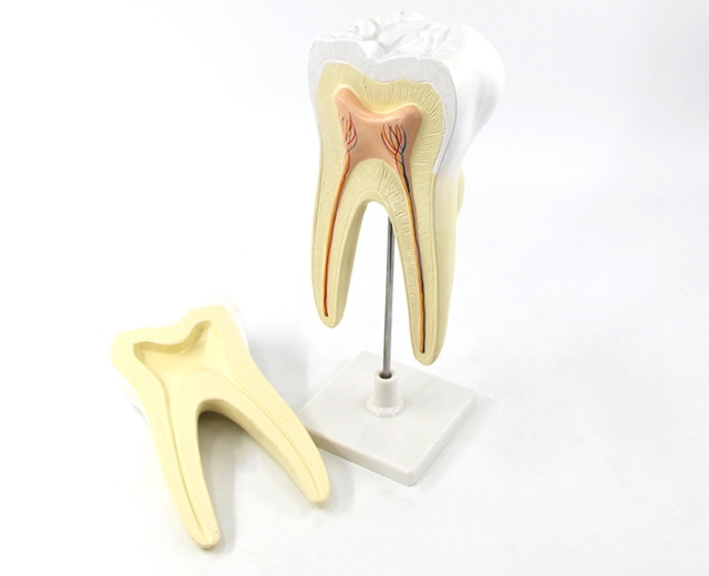
Human molars anatomy model, kindergarten teeth teaching AIDS, large teeth model with base
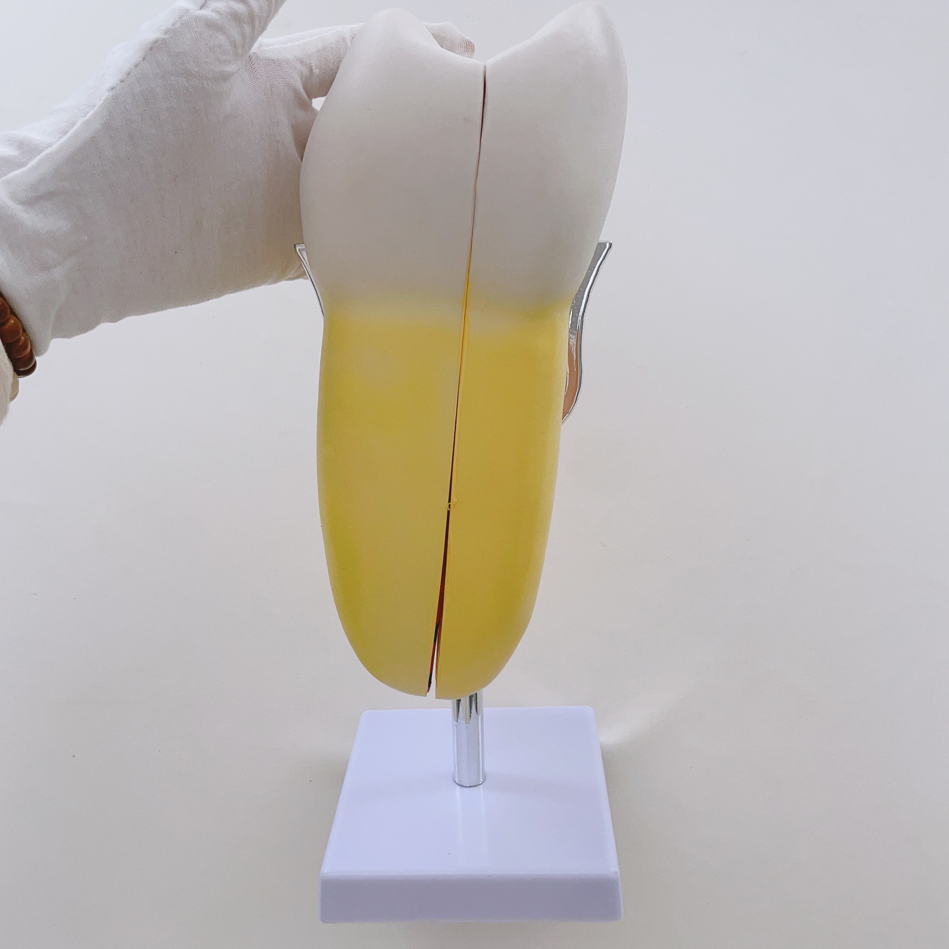
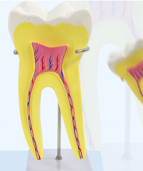
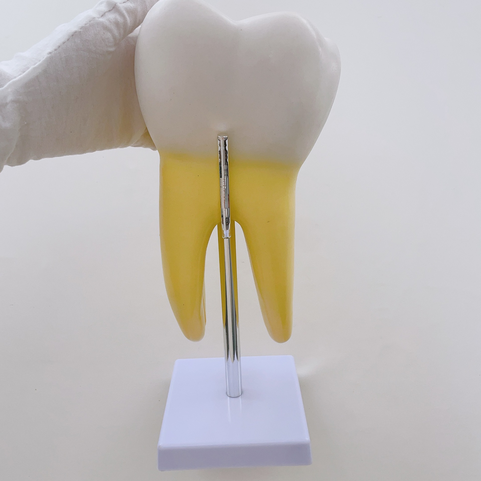
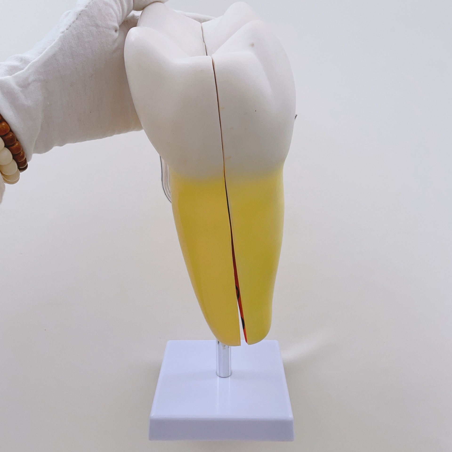
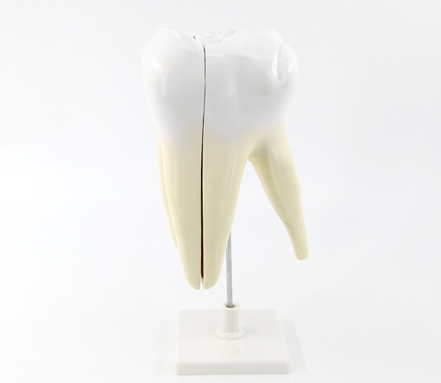
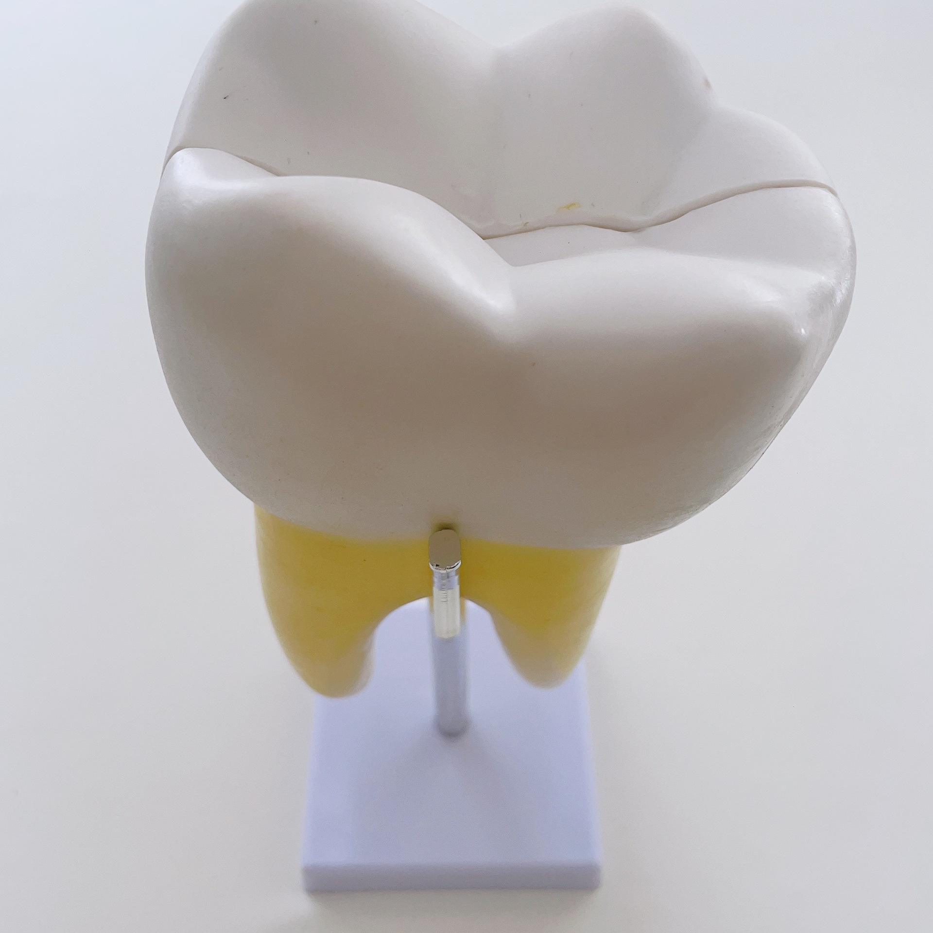
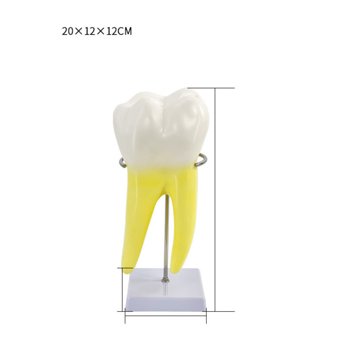
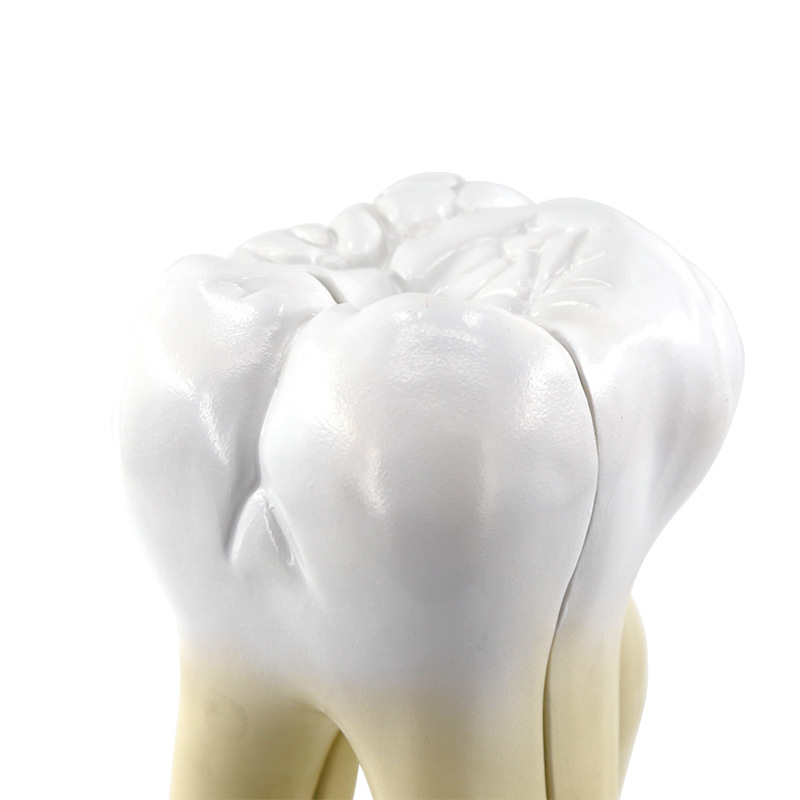
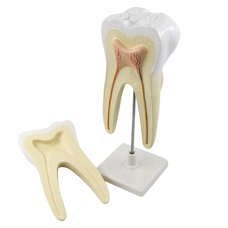
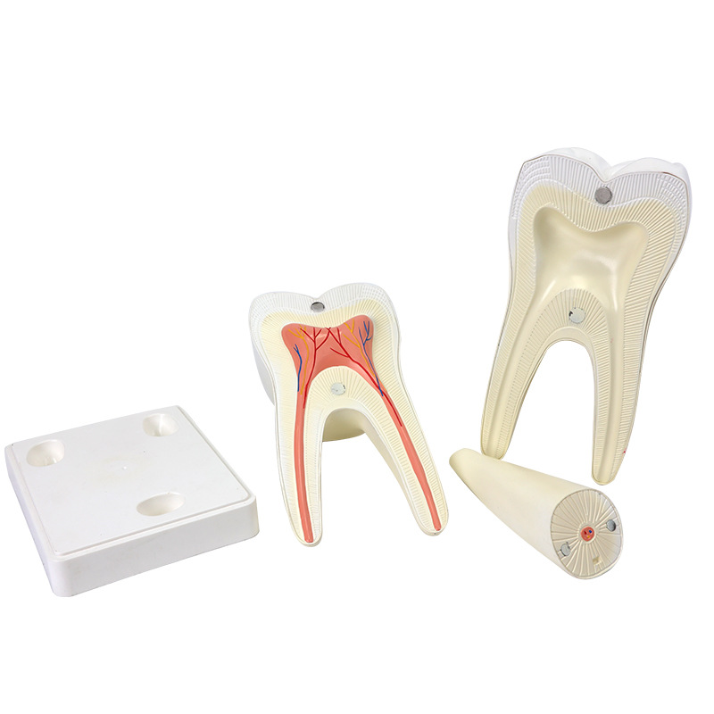
This model features a 6 – fold magnification of molars, consisting of 2 parts. It is designed for educational purposes, enabling students and professionals to observe the intricate anatomical structures of molars in detail. Ideal for dental education settings, it provides a clear and enlarged view of molar features, facilitating a better understanding of molar anatomy.
Product application
1.Dental Education
In dental schools, this model serves as an essential teaching aid. It helps students learn about molar anatomy, such as the structure of enamel, dentin, pulp cavity, and root canals.The 6 – fold magnification allows students to observe fine details that are difficult to see on real – sized teeth, enhancing their understanding of molar morphology and preparing them for clinical practice.
2. Training for Dental Professionals
For dentists, dental hygienists, and other dental professionals, this model can be used for continuing education and training.It enables them to review molar anatomy, study the progression of dental diseases like decay in relation to molar structure, and practice procedures such as filling placement and root canal treatment in a simulated environment.
3.Patient Education
In dental clinics, this model can be employed to educate patients.It helps dentists explain molar – related dental issues, such as the causes and consequences of tooth decay, the importance of proper oral hygiene for molar health, and the steps involved in various dental treatments.The enlarged view makes it easier for patients to visualize and understand these concepts.
4. Research and Development
In dental research institutions, the model can be used as a reference for studies related to molar development, dental materials testing, and the evaluation of new dental treatment techniques.Researchers can use it to compare the effects of different substances or procedures on molar anatomy in a controlled and observable manner.