
# Human Duodenal Anatomy Teaching Model – A Precise Teaching Aid Solution for Medical Education
I. Product Overview
This human duodenal anatomy teaching model strictly adheres to human anatomy standards, accurately presenting the anatomical structure of the duodenum and its adjacent organs such as the liver, gallbladder, and pancreas. It provides a highly realistic and detachable teaching tool for medical education, clinical demonstration, and anatomical research, assisting professionals in deeply analyzing the anatomical logic and pathological connections of the digestive system.
Ii. Core Values
(1) Breakthrough in anatomical accuracy
Relying on human cross-sectional anatomical data and 3D modeling technology, the model accurately reproduces the morphological characteristics of the duodenal bulb, descending part, horizontal part and ascending part, and clearly presents the microscopic structures such as the duodenal papilla and circular folds. The course of the portal vein, hepatic artery and common bile duct within the hepatoduodenal ligament, as well as their adjacent relationship with the pancreatic head, are all replicated 1:1, providing a “gold standard” reference for the teaching of digestive system anatomy.
(2) Modular Teaching Adaptation
It adopts a multi-component detachable design, allowing each section of the liver, gallbladder, pancreas and duodenum to be independently disassembled and combined. It supports step-by-step teaching from local anatomy (such as separately showing the descending part of the duodenum and the opening of the pancreatic duct) to systematic association (fully presenting the liver-biliary-pancreaticoduodenal pathway), and is suitable for various scenarios such as basic anatomy teaching and training in the diagnosis and treatment of digestive system diseases, helping trainees build a three-dimensional knowledge system of “macroscopic – microscopic” and “local – systematic”.
(3) Professional material guarantee
It is made of medical-grade polymer composite materials, featuring a biomimetic texture of tissues and a color that restores the physiological color of human organs. It is not prone to oxidation or deformation over long-term use. The base adopts a stainless steel bracket and high-density resin to ensure the stability of the model. It is suitable for high-frequency usage scenarios such as medical college laboratories and clinical skills training centers, providing long-lasting and reliable hardware support for teaching demonstrations and practical training.
Iii. Application Scenarios
- ** Medical Education System ** : In the anatomy courses of medical colleges and universities, it serves as a visual teaching aid for theoretical teaching to assist teachers in explaining the key points of duodenal anatomy; In the laboratory class, students are provided with practical exercises to disassemble and identify structures, thereby strengthening their memory of anatomical knowledge.
- ** Clinical training scenarios ** : In specialized training programs such as gastroenterology and general surgery, it is used to analyze the anatomical basis of diseases like duodenal ulcers and perampullary cancer, and to assist in the construction of clinical thinking; Before the surgical simulation training, help the surgeons familiarize themselves with the anatomical layers of the surgical area.
- ** Medical Science Popularization Promotion ** : In hospital health management centers and medical science popularization exhibition halls, knowledge about digestive system health is explained to patients and the public in an intuitive way, facilitating the development of disease prevention and health management science popularization work.
This model takes anatomical accuracy as the foundation and teaching practicality as the orientation, providing professional teaching aid support for all links of medical education, helping to cultivate high-quality medical talents, and promoting the deep integration of digestive system anatomy teaching and clinical practice.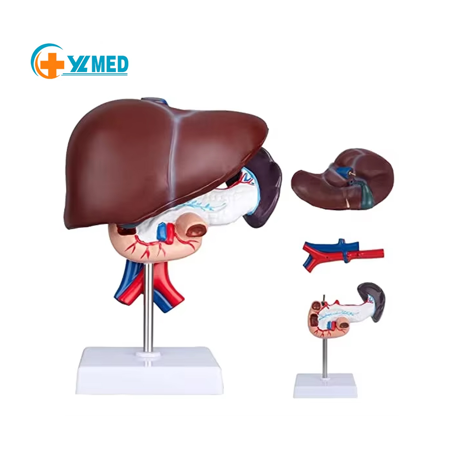
Post time: Jun-06-2025