
Exquisite craftsmanship, ultimate restoration
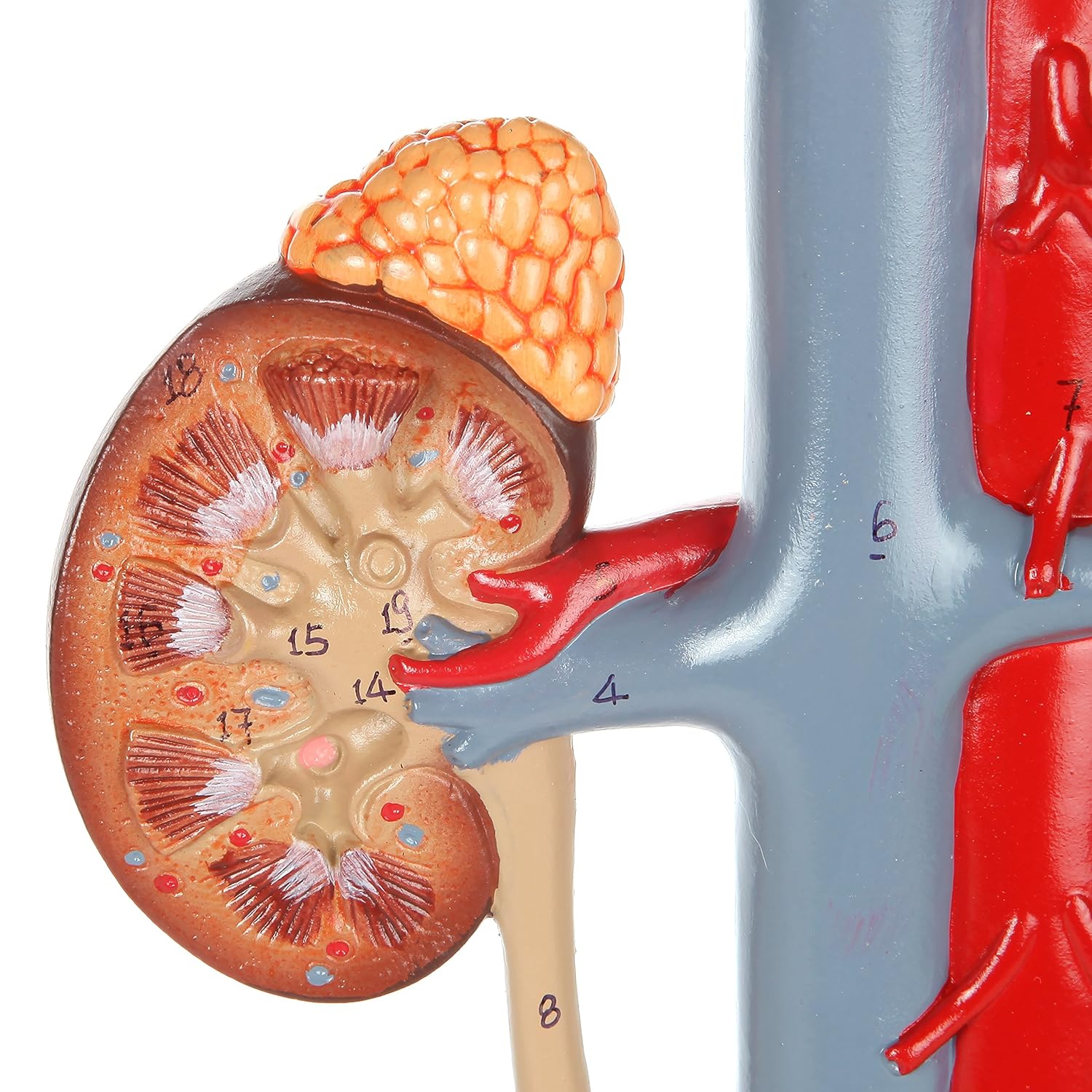
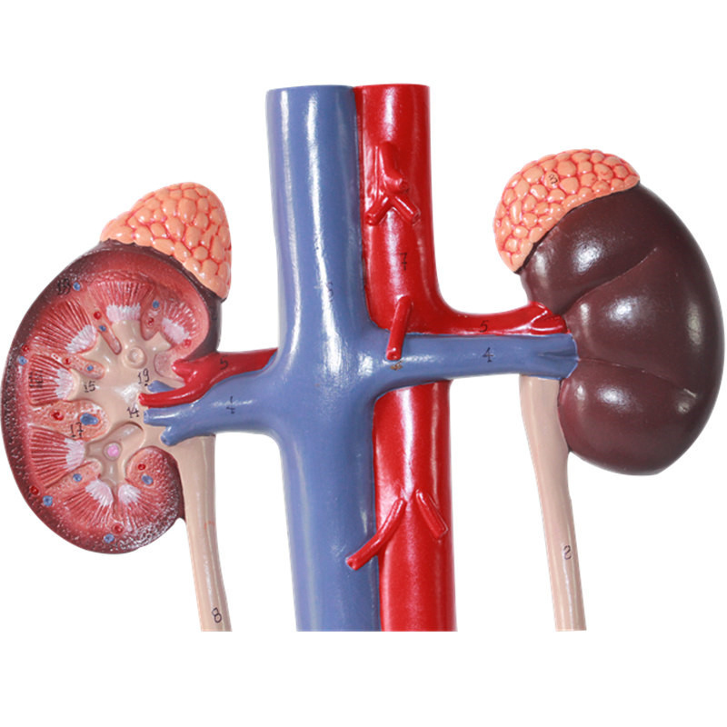
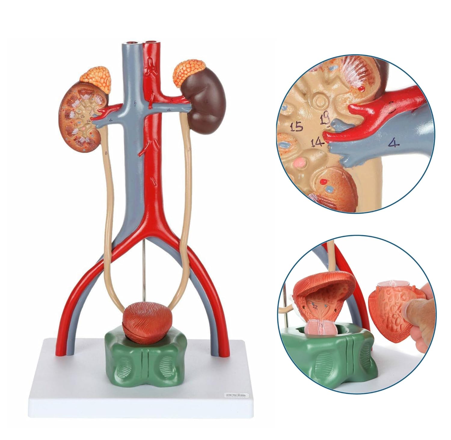
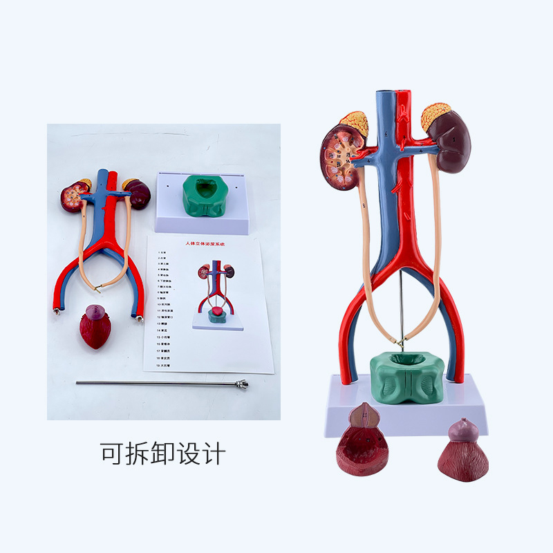
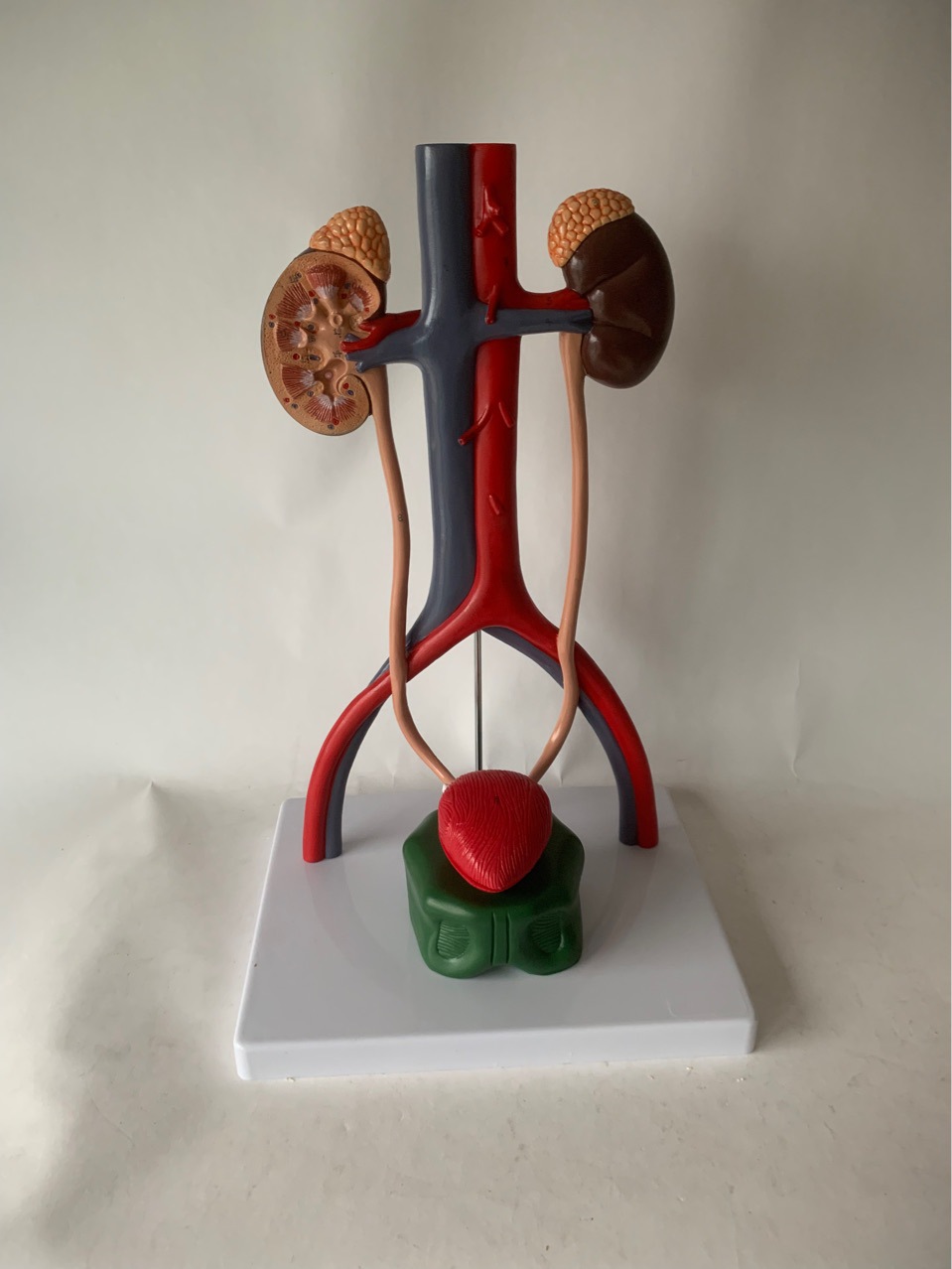
The human urinary system anatomical model is crafted with meticulous precision, achieving an ultimate restoration of the human urinary system. The kidney section in the model allows for a clear view of the densely packed renal corpuscles in the renal cortex, as well as the striated renal tubules in the renal medulla, as if presenting the microscopic structure of a real kidney in miniature. The shapes, positions, and connections of the ureters, bladder, and major blood vessels are also highly consistent with the actual human condition. Every detail has been rigorously examined and precisely shaped, providing visitors with a stunning visual experience.
## High-quality Materials, Long-lasting Durability
In terms of material selection, this model uses high-quality PVC material. This material is not only tough in texture, capable of withstanding frequent handling and display, but also has excellent anti-aging and anti-wear properties. Even after long-term use, the color and structure of the model will not show significant changes, always maintaining its original perfect state. This feature is undoubtedly a great advantage for medical exhibitions where frequent display and use are required, as well as for long-term use in medical teaching and research.
## Educational Value Is Outstanding, Facilitating Medical Development
This model holds particularly significant value in the field of medical education. For students at medical schools, after learning the theoretical knowledge of the urinary system in class, by observing and studying this anatomical model, they can transform abstract knowledge into intuitive understanding, significantly enhancing their learning outcomes. For instance, when understanding the process of urine formation and excretion, students can clearly see through the model how blood flows through the kidneys, undergoes filtration in the renal units, forms urine, and then enters the bladder for storage before being excreted out of the body.
For medical professionals, this model can serve as an important auxiliary tool for clinical teaching and case discussions. When explaining diseases related to the urinary system, such as kidney stones and bladder inflammation, it can accurately indicate the affected areas, analyze the causes and effects of the diseases, and assist medical staff in formulating better treatment plans.
Furthermore, at this medical exhibition where the latest technologies and products in the industry are showcased, the appearance of the human urinary system anatomical model provided an opportunity for related enterprises and research institutions to communicate and collaborate. It demonstrated [Company Name]‘s strength and innovation capabilities in medical education product research and development, and is expected to drive further development of anatomical model technologies and applications in the entire medical education industry.
With the increasing demand in the medical field for precise education and visual learning, the launch of this human urinary system anatomy model is timely. It not only adds a bright touch to this medical exhibition, but will also play a significant role in future medical education and medical practice, helping to cultivate more outstanding medical talents and promoting the continuous progress of the medical industry.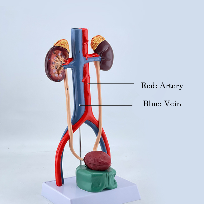
Post time: Aug-29-2025