
# Shocking Release of Foot Anatomy Model, Facilitating New Breakthroughs in Medical Education
### 1. Precise Reproduction, Every Detail of Anatomy Clearly Revealed
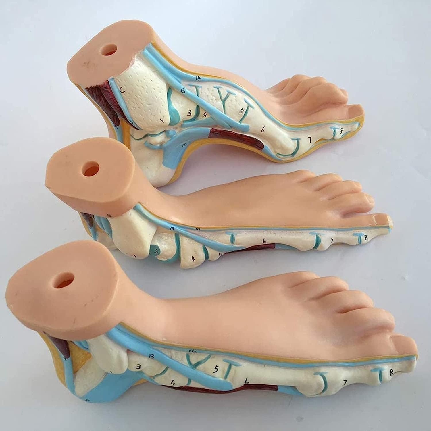
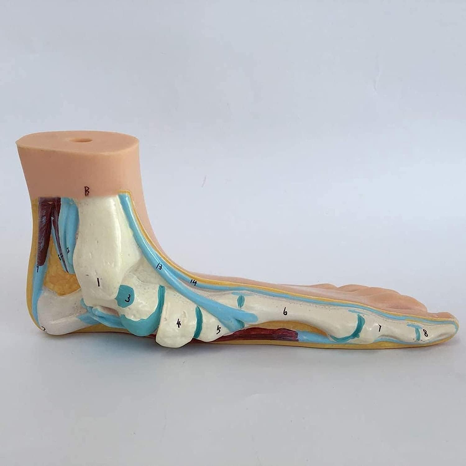
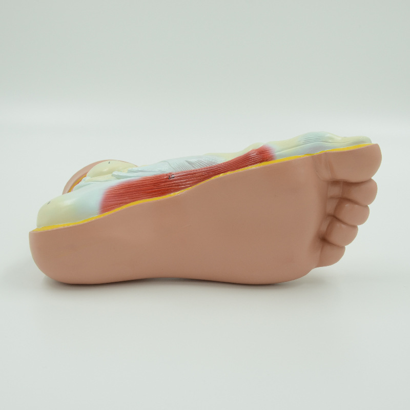
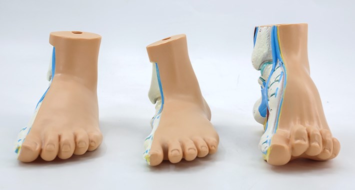
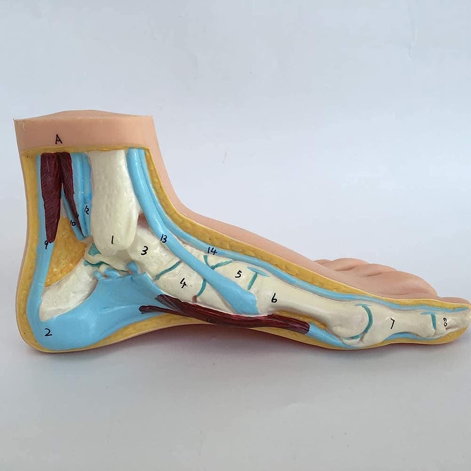
This foot anatomy model has meticulously replicated the physiological structure of the foot. From the perspective of bones, the shape, size, and joint surface texture of the foot bones are highly consistent with those of the real human body. The concave and convex shapes of the talus bones, the thickness variations of the metatarsal bones, and even the subtle curvatures of the phalanges are all calibrated by medical experts against human specimens, clearly presenting the mechanical support structure of the foot bones. In terms of muscle tissue, based on the human anatomy atlas, the muscle distribution layers are precisely restored. The thickness differences of the plantar muscles, the extension direction of the muscle groups in the lower leg to the tendon of the foot, and even the simulation of the shape during muscle contraction, are all lifelike, enabling learners to intuitively understand how muscles work together to control foot movements. The nervous and vascular systems are even more meticulous. The branching directions of nerves, the connection structures of blood vessels, small details such as the shape of the arch of the foot artery and the shallow position of the cutaneous nerves, are all clearly distinguishable, completely presenting the complex interconnection of the foot’s nerve and vascular network, providing an intuitive carrier for explaining knowledge such as foot sensation conduction and blood circulation.
### 2. Multi-scenario Adaptability, Comprehensive Support for Teaching Practice
In medical school classrooms, it serves as a “visual assistant” for theoretical knowledge. When teachers explain the chapter on foot anatomy, they can use the model to split and display, from the overall structure to the local details, analyzing the combination relationship of bones, muscles, nerves, and blood vessels layer by layer, enabling students to break free from abstract textual descriptions and quickly establish spatial cognition, understanding the anatomical basis of the foot as a movement and load-bearing organ. In the clinical doctor training scenario, the model becomes the “pathological simulation platform” for pathological analysis. When dealing with common foot diseases such as fractures, tendinitis, and nerve compression syndromes, the model can simulate the lesion location, analyze how bone displacement compresses nerves and blood vessels, and how muscle damage affects the movement function of the foot, helping doctors understand the pathogenesis from the anatomical perspective and assist in formulating treatment plans. Even in rehabilitation medicine teaching, the model can also play a role, used to demonstrate the rehabilitation training principles after foot injury, explaining how muscle strength recovery and joint range of motion training can improve foot function, becoming an important teaching aid that connects basic medicine with clinical practice.
“We are committed to providing high-quality teaching aids for medical education. The launch of this foot anatomy model is a deep response to the teaching needs.” [Company Name]‘s director stated that they hope to break the communication barrier between theory and practice through advanced anatomical models, making medical learning more efficient and intuitive. Currently, this model is available for reservation on the independent website, attracting inquiries and orders from numerous medical institutions and educators. It is expected to become a new favorite in medical teaching scenarios and drive the advancement of foot medical teaching to a new level.
Post time: Aug-27-2025