
For medical education Human eye model Sensory model orbital model double magnification anatomy

For medical education Human eye model Sensory model orbital model double magnification anatomy

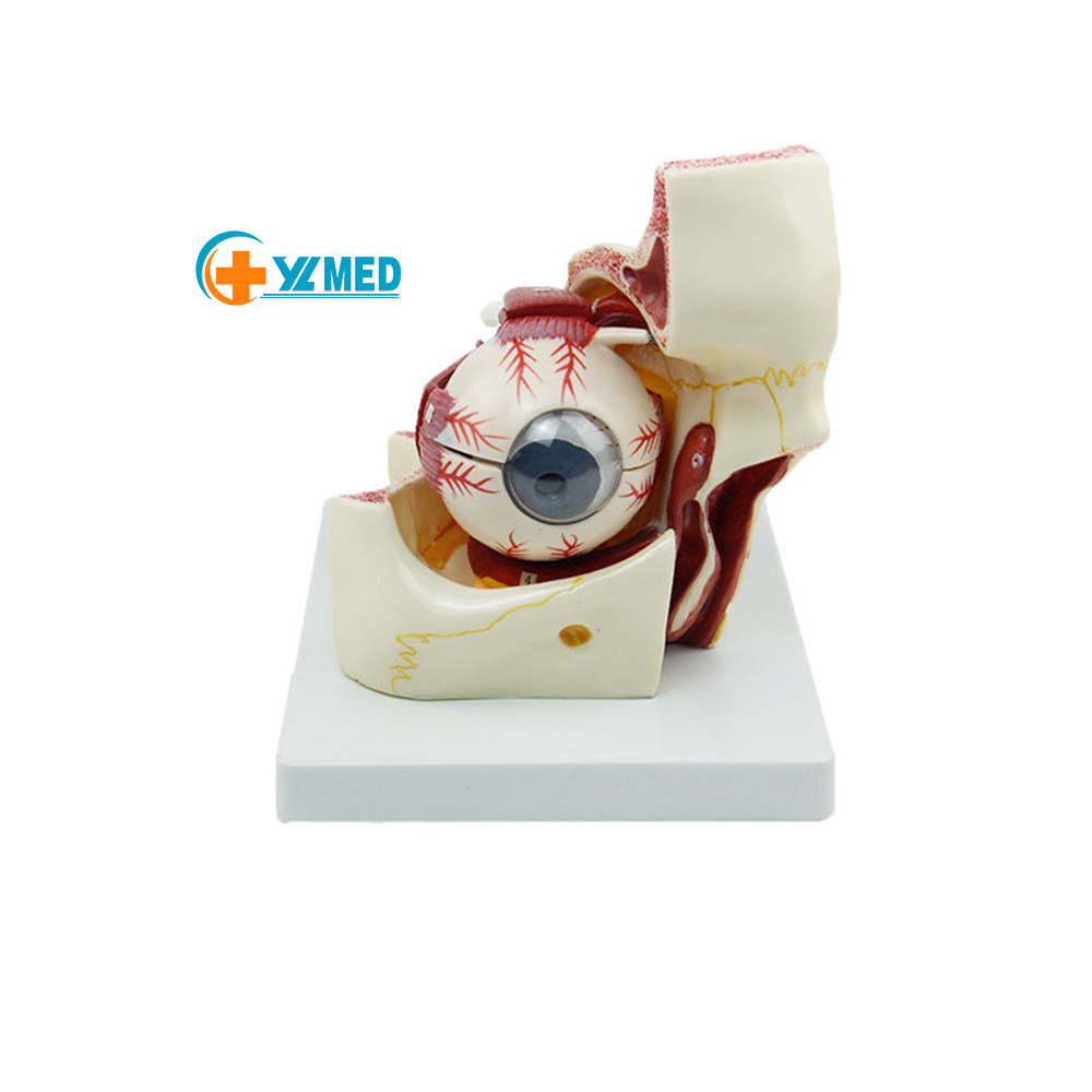
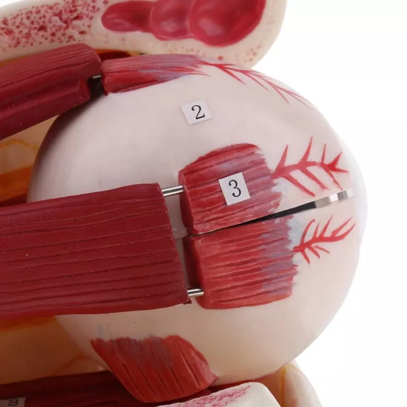
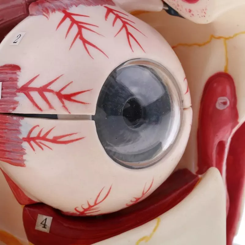
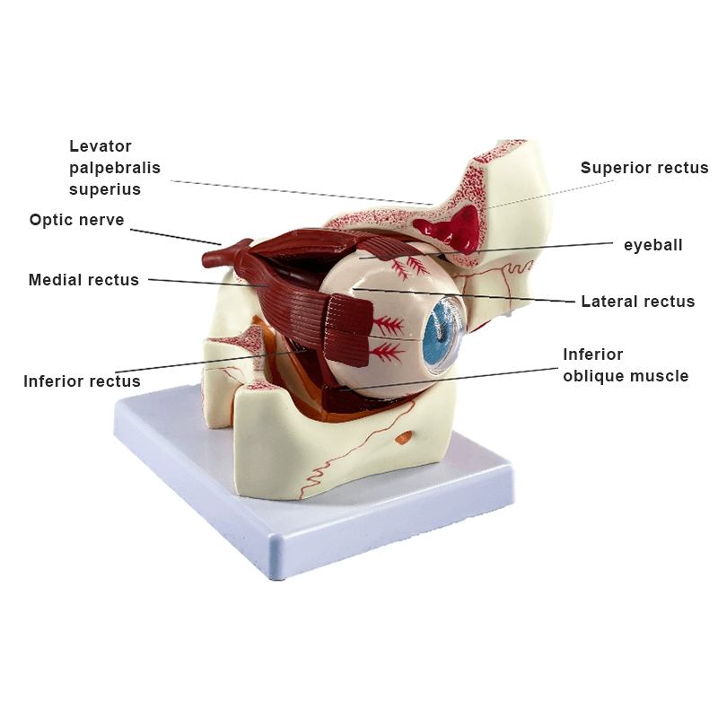
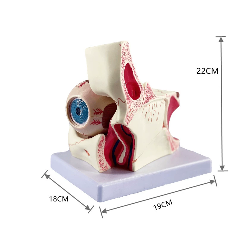
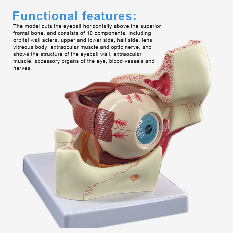
1. In the model, the eyeball was cut horizontally above the maxilla, and was composed of 10 components, including orbit, ocular wall sclera, upper and lower half, lens, vitreous body, extraocular muscle and optic nerve; 2. Display the corpuscular wall of the eye (sclera, cornea, iris, ciliary body, choroid and retina), the contents of the eye,extraocular muscles, accessory organs of the eye, blood vessels and nerves and other structures; 3. Triple magnification.
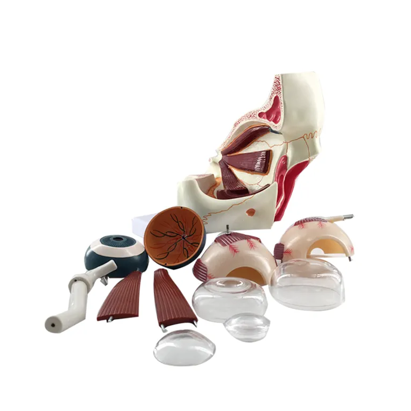
1. Detachable eyeball design
Can look at the eye hierarchy, conducive to teaching
Can look at the eye hierarchy, conducive to teaching
2. Eco-friendly PVC material
A widely used synthetic material with non-flammability
3. Clear details for easy observation
The model is suitable for medical teaching/training explanation/decoration/doctor-patient communication
 、
、
Product Advantages:
1. Visual teaching AIDS are detailed and realistic
The cutting surface is clear and vivid, with digital marks, which is conducive to teaching display and medical students’understanding;
2. Environmental protection material computer color matching
Environmental protection PVC material, computer color matching, manual printing, long service life
durable.
3. The model can be disassembled and assembled
When teaching physiological health courses in schools, it is used as visual teaching AIDS to show.
Products categories
-
Advanced Medical science muscle Intramuscular i...
-

85cm movable miniature human skeleton model for...
-
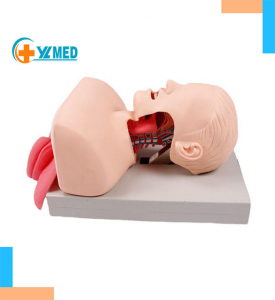
Adult Electronic Tracheal Intubation Teaching M...
-
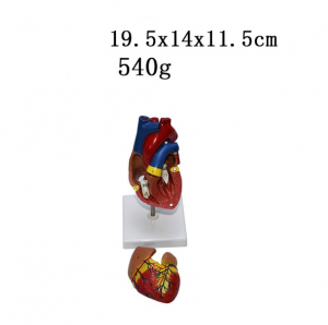
Medical Human Cardiac Anatomy Models Education ...
-
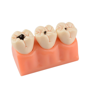
Dental Caries Teeth Model 4 Times Dental Tooth ...
-
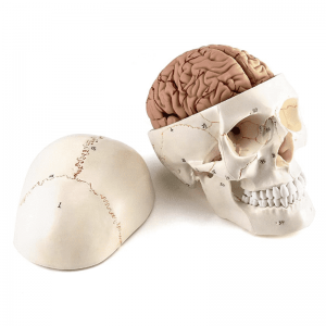
Anatomical models of the human skull and brain ...