Product Detail
Product Tags
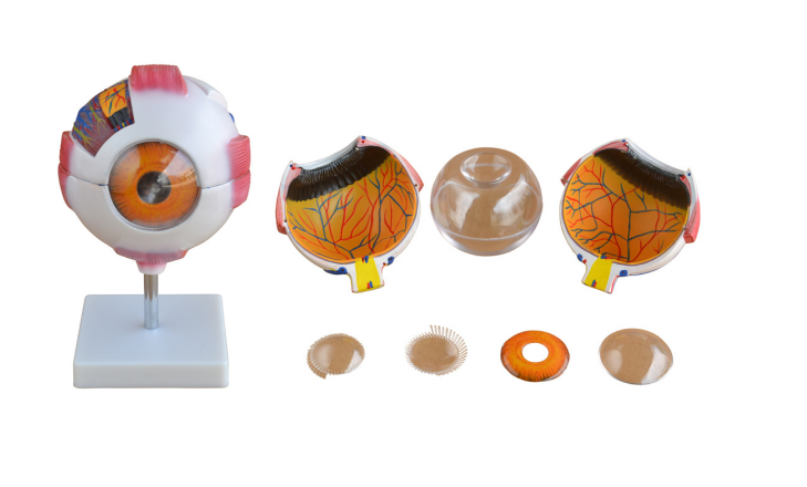
- ▲6X Enlarged Eye Model – The 6-parts eyeball model is medical level and shows the 12 eyeball positions: Corneal, Sclera, Choroid, Retina, Iris, Lens, Vitreous Body, Optic Nerve, Central Fovea, Cochlear Vein, Ciliary Body, Central Retinal Arteriovenous.
- ▲Colorful Hand Painted Craftsmanship – The eye model is detailed and hand painted with fine craftsmanship. Different parts of the epidermis model are marked with different numbers, which is convenient for accurate teaching and display.
- ▲Premium Material – The 3d model of the eyeball is made of non-toxic environmentally friendly PVC material, easy to clean and will last for years. It also has a durable base for public explanation and demonstration.
- ▲Versatile Application – The human anatomical eye model is suitable for doctor-patient communication. It can also be used as a teaching and study tool for ophthalmologists, medical school students, practitioners, health care professionals, schools and universities and so on.
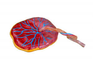
- This model was used to show the anatomical structure of the human eyeball, such as the three layers of membranes (outer membrane, media and intima) of the eyeball wall and the main refractive body, lens and vitreous body filling the interior. The model was made of PVC, magnified 6 times according to the real object, and placed on the base.
Size: 12x12x28CM
Packing: 18pcs/carton, 53x39x55cm, 22kgs
Previous:
Digestive system model
Next:
Magnifying model of eyeball and orbit