Medical Science Teaching Model PVC Normal VS Asthmatic Trachea Comparison Model for Respiratory Education
Medical Science Teaching Model PVC Normal VS Asthmatic Trachea Comparison Model for Respiratory Education
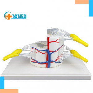
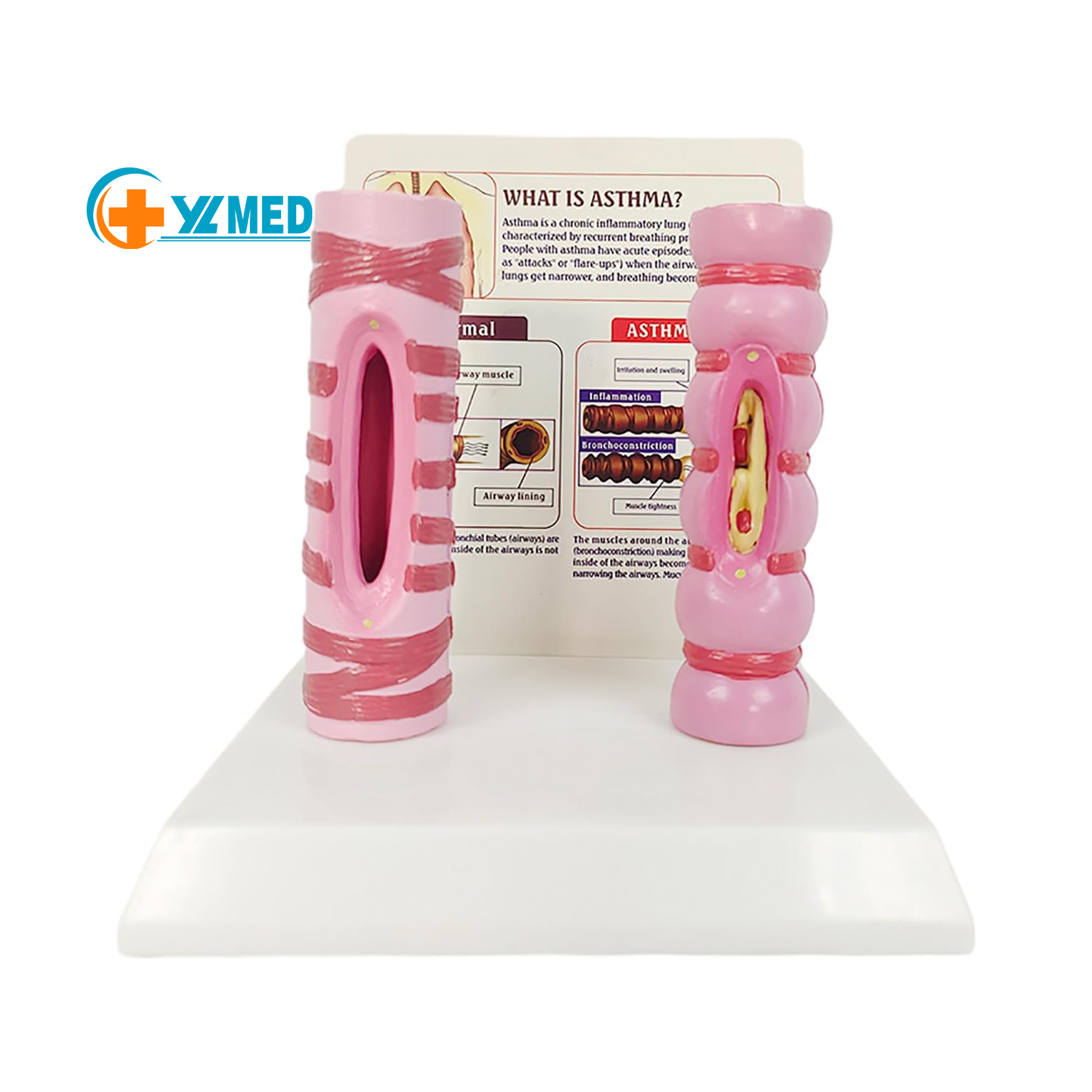
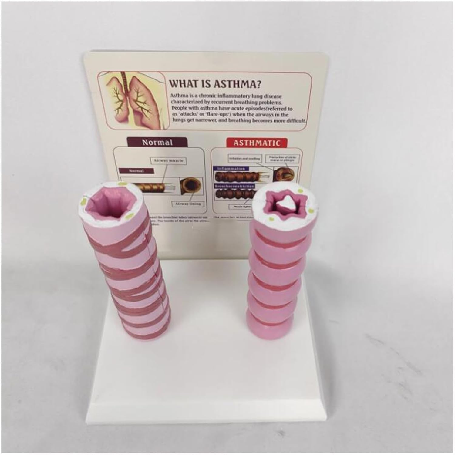
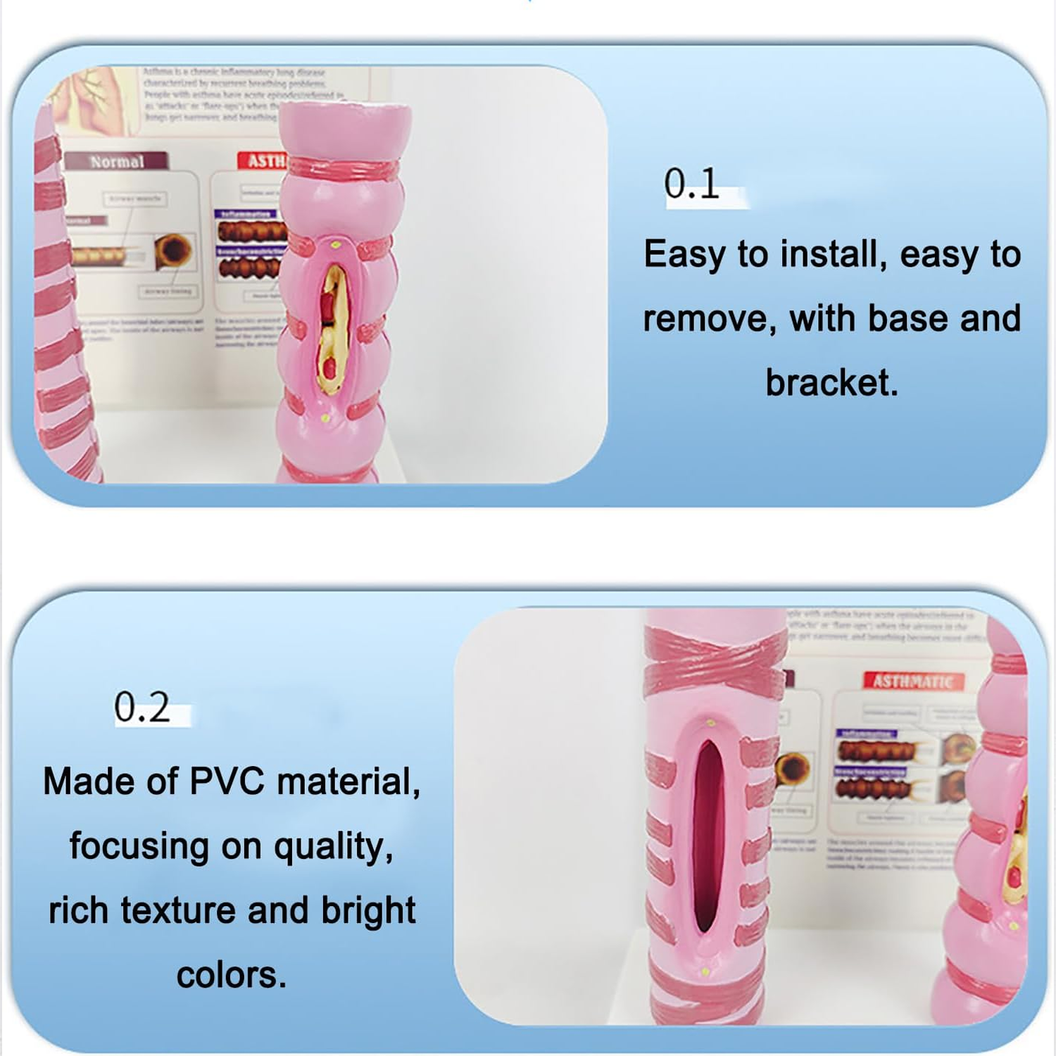
1. Made of environmentally friendly PVC material, simulating the airway muscles and mucosal layer structure of the trachea, with clear color layers (smooth texture of normal trachea, swelling and mucus details of asthmatic trachea), durable and odorless, suitable for long-term teaching display.
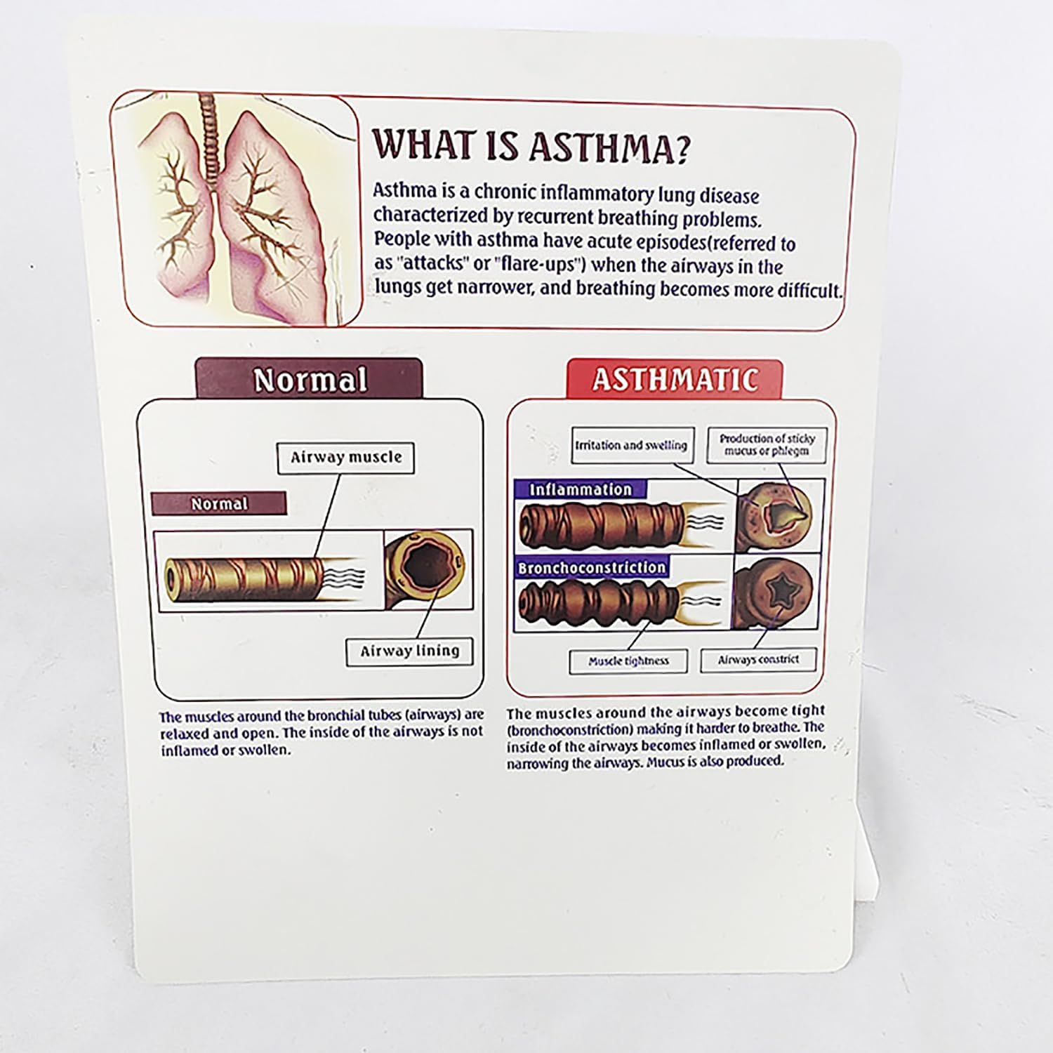
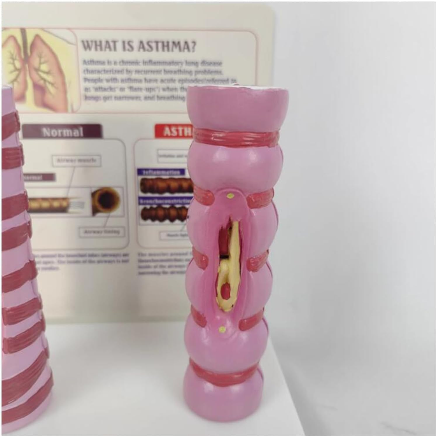
2. Includes two comparable models of normal trachea and asthmatic pathological trachea, with an explanatory display board clearly marking the anatomical differences of airway muscles and mucosal layers. The structure is stable and the comparative display is intuitive.
3. Mature supply chain, sufficient inventory, supporting bulk procurement, fast delivery after ordering, ensuring teaching and research progress.
4. The model and display board are easy to install, ready for teaching explanation out of the box, no complicated assembly
required, easily used by medical staff, teachers, and students.

The dual-model comparison design can intuitively present the anatomical differences between normal and asthmatic trachea, combined with the pathological mechanism analysis of the explanatory display board. Compared with ordinary respiratory system models, it has stronger teaching pertinence and clearer pathological cognition, helping learners quickly understand the airway lesion principle of asthma, and is an efficient tool for respiratory disease teaching.

Suitable for respiratory anatomy course teaching in medical colleges, clinical training in hospital pulmonology departments, airway lesion research in asthma research institutions, and intuitive explanation of asthma knowledge in medical science popularization. It is a practical tool for carrying out respiratory anatomy teaching, asthma pathological analysis, and health science popularization.

Place the two models and the display board on the teaching table to intuitively compare the morphological differences between normal and asthmatic trachea; combined with the text on the display board, explain the pathological changes of airway muscles and mucosal layers. During teaching, hold the model to point out anatomical features; in scientific research, observe lesion details from multiple angles. After use, clean the surface of the model and store it in a dry place.