Medical teaching Human upper limb muscle Detachable biceps anatomical model with vascular nerve human arm model

Medical teaching Human upper limb muscle Detachable biceps anatomical model with vascular nerve human arm model
Product Description
Human upper limb muscle detachable biceps anatomy model with blood vessels and nerves human arm model

Description
* Details:
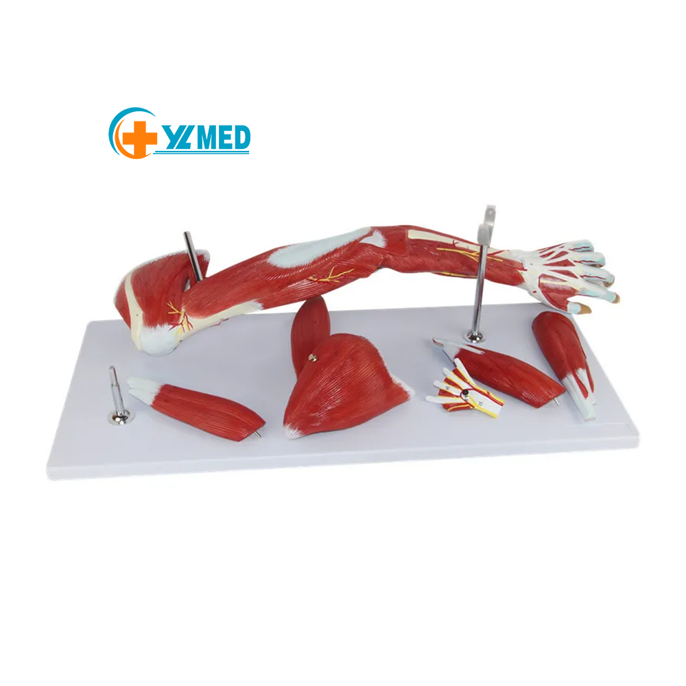
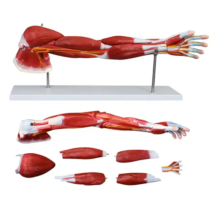
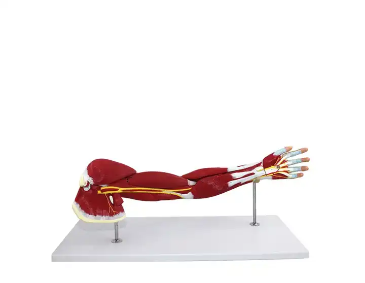
Anatomical model of upper limb muscle The model was composed of seven parts, including upper limb muscles, deltoid muscle, triceps brachii, radial brachialis, pronator teres, flexor digitorum superficialis, brachial plexus and axillary artery. It showed the structures of upper limb belting muscle, brachial muscle, anterior group of forearm muscle, posterior group of forearm muscle and hand muscle, with a total of 87 site indicators.
Detailed Images
|
Specification
* Material:PVC
* Process: Computer matching advanced color painting * Size :73*23*10.5cm * Packing:77.5*33*23 cm;1 pcs/ctn;6 kg. |
Human upper limb muscle detachable biceps anatomy model with blood vessels and nerves human arm model


|
Structural advantage
1. This model shows 7 components, including the superficial muscle of the left arm with the shoulder, the deep muscle, the upper limb muscle, the deltoid muscle, the triceps of the brain, the burning muscle of the brain, the teres pronator, the flexor digitalis superficial, the brachial plexus and the axillary artery
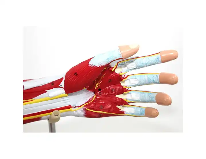
2.These7 parts are detachable for further careful study; 3. Accurate reproduction of muscle, blood vessel, nerve and bone components, can clearly understand the upper limb band muscle,arm muscle, forearm muscle anterior group, posterior group and hand muscle structure; 4. The professional design of the palmar longus can be split, showing the hand muscle stratification hand bone, ligament, muscular ciliary and its main vessels and nerves and other structures; 5. There are digital signs and corresponding text descriptions for the convenience of teaching. |