
Ultrasound-guided Central Venipuncture Training Model Can Be Used for Internal Jugular Venipuncture and Subclavian Venipuncture
Ultrasound-guided Central Venipuncture Training Model Can Be Used for Internal Jugular Venipuncture and Subclavian Venipuncture
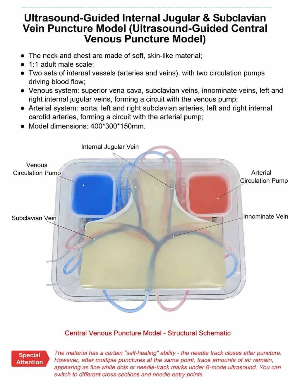
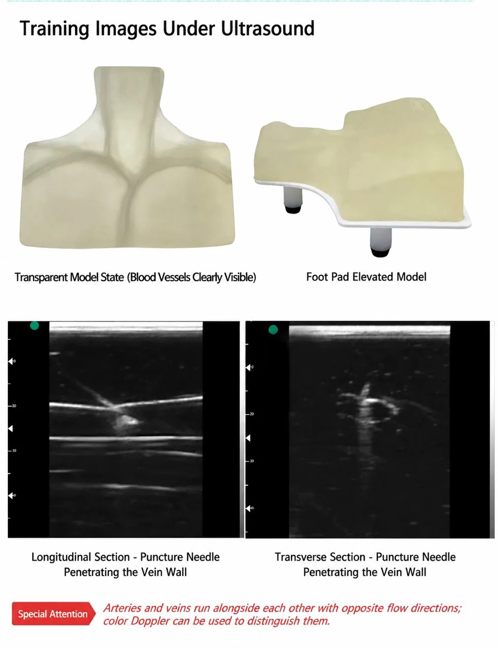
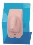
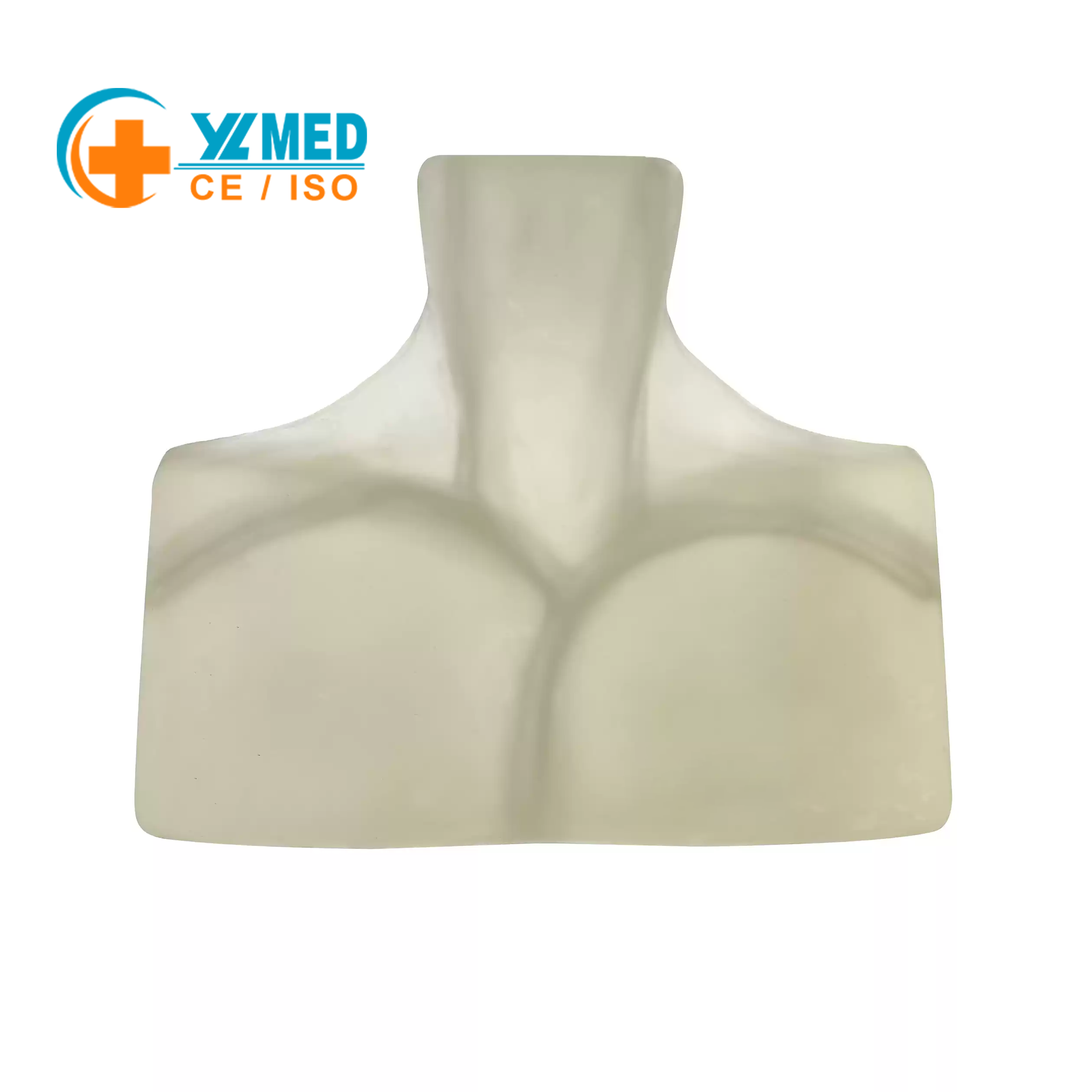
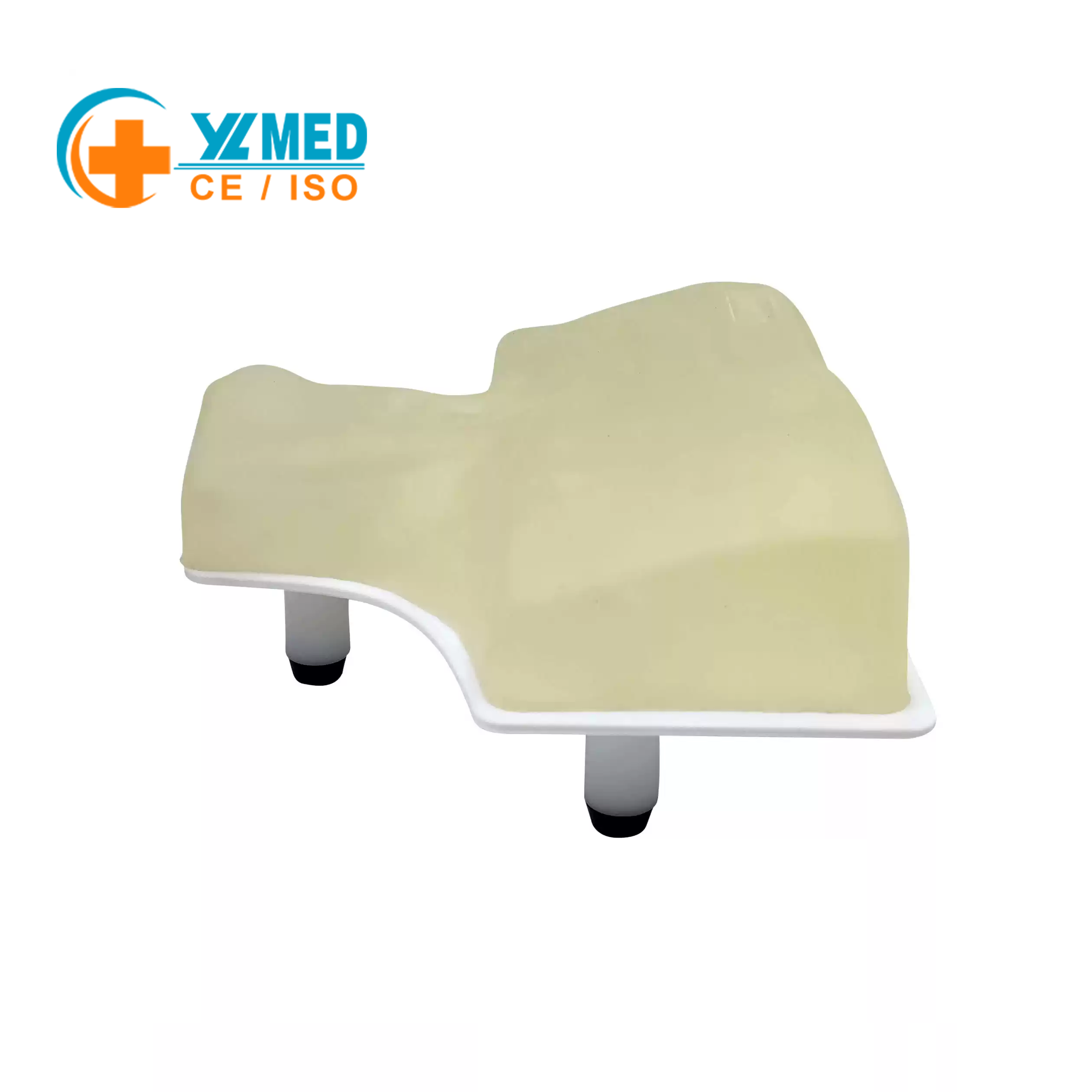
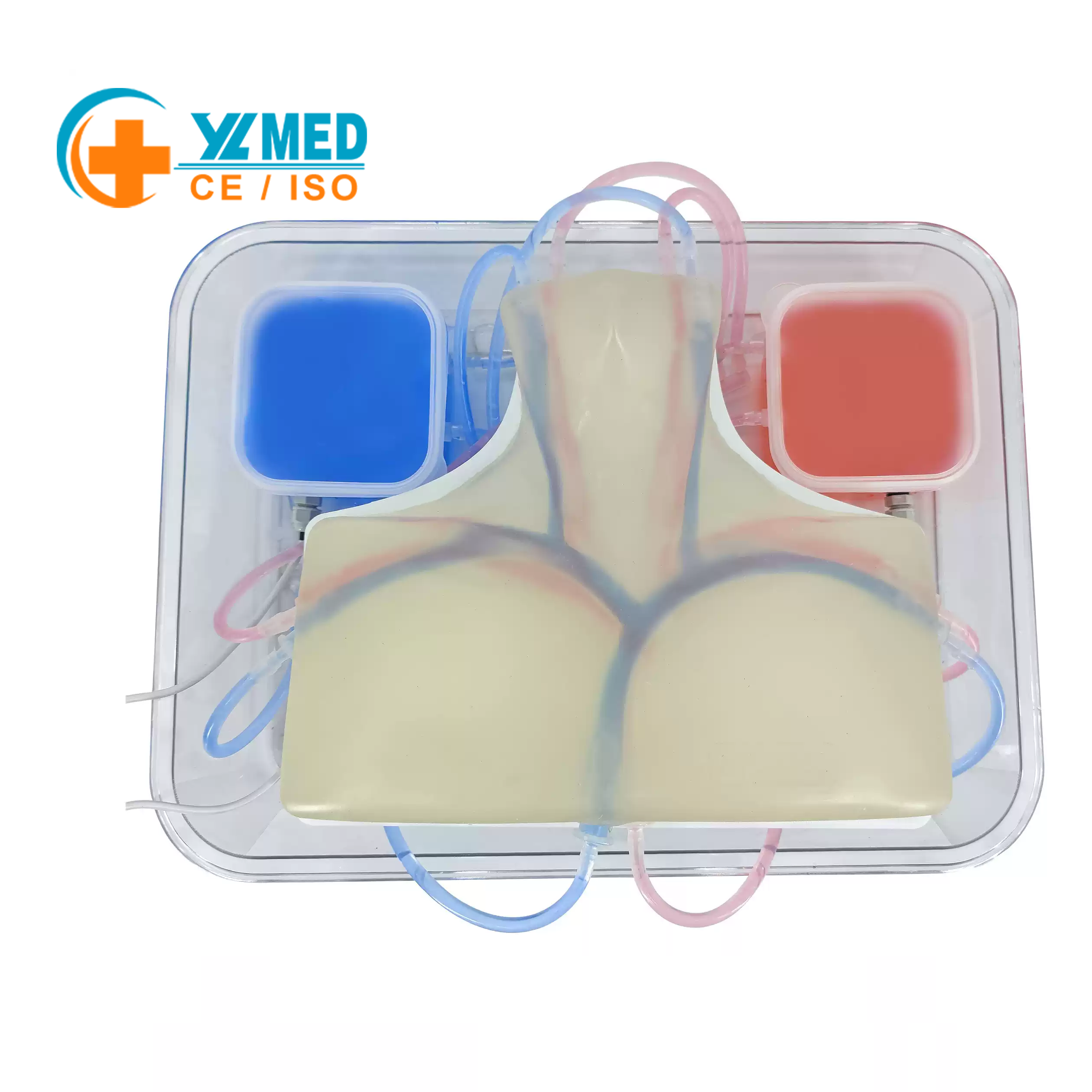
Accurately restores the anatomical structure of cervical blood vessels, including vascular and tissue modules that can simulate ultrasound imaging. The transparent design intuitively presents the spatial relationship between the puncture needle and blood vessels, helping learners master core skills of ultrasound-guided central venous puncture.
Supports the simulation of ultrasound-guided central venous puncture operations. The vascular imaging and puncture needle position can be observed through an ultrasound device (to be equipped separately), concretizing abstract interventional operations and improving the learning efficiency of puncture skills.
The operation is close to clinical practice. Combined with an ultrasound device, it can simulate the real process of “ultrasound localization – puncture needle insertion – catheter placement”. The transparent structure is convenient for teachers to guide and students to self-correct, suitable for interventional medicine classroom training and clinical pre-operative simulation.