
Biology Science Education Equipment Veterinary Medical Simulation Animal Dog Anatomy Model Canine Eye Model
Biology Science Education Equipment Veterinary Medical Simulation Animal Dog Anatomy Model Canine Eye Model
F (Feature)
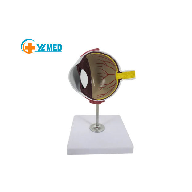
This is a canine eye anatomy model made of high-quality fiberglass, sized at 12×5×10cm. It features a detailed cross-sectional view of the dog eye, showcasing structures like the cornea, lens, retina, blood vessels, and optic nerve, mounted on a stable base.
A (Advantage)
The model provides a clear, magnified view of the canine eye’s internal anatomy, making it easy to identify and study key
structures. Its durable fiberglass construction ensures long-term use in educational and clinical settings.
B (Benefit)
For veterinary students, educators, and clinic staff, this model is an essential tool for teaching and learning canine ophthalmic anatomy. It helps students visualize eye structures, understand their functions, and prepare for clinical examinations and treatments.
C (Conclusion)
A compact, durable, and highly detailed canine eye model, ideal for veterinary schools, animal clinics, and medical education programs. Contact us today to learn more about bulk purchasing options.
Product Description
Biology Science Education Equipment Veterinary Medical Simulation Animal Dog Anatomy Model Canine Eye Model
Showcasing the eye structure of dog, suitable for veterinary, clinic, and medical schools, made of high-quality fiberglass.
|
Product Name
|
Canine Eye Model
|
|
Product Size
|
12*5*10cm
|
|
Function
|
Students Understand Animals Structure
|
|
Instructions
|
English
|

Detailed Images
Biology Science Education Equipment Veterinary Medical Simulation Animal Dog Anatomy Model Canine Eye Model

Products categories
-

Medical Science 5x Life Size Vertebral Partial ...
-
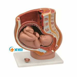
Human Female Pelvic Section Pregnancy Anatomica...
-
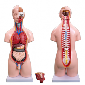
A total of 20 55CM male and female torso models...
-
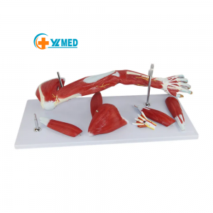
Medical teaching Human upper limb muscle Detach...
-
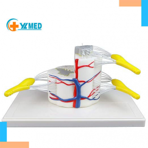
Medical Science YULIN YL-235 5x Life Size Verte...
-
Arterial vein blood vessel dissection model hum...